Preparation with CBCT crucial to maxillary implant achievement
- February 17, 2016
- Feature
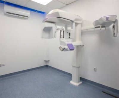
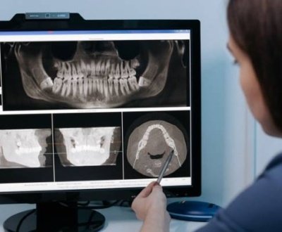
As more and more dentists are carrying out implant placement, a fresh investigation done by researchers from the University of Texas School of Dentistry at Houston, discovers that vigilant management preparation with cone-beam computed tomography is decisive for the achievement of instantaneous implant location, particularly at the lateral incisor area on account of restricted accessibility of alveolar bone.
In harmony with the investigators, the realization of implant management is contingent on facts and figures on the stature, distance across, morphology, and compactness of alveolar bone adjacent to the implant site. Although orthodox radiographic practices had earlier been reflected the benchmark for treatment preparation, yet the correctness of these practices is bargained by imaging falsification and overlying.
Evaluation Prior to the Operation
The researchers observed that the general alveolar measurement and shape at anterior maxilla have not been completely evaluated. In the present investigation, the investigators established that in the patient regiment the usual alveolar measurement at the anterior maxilla is roughly 18.83 mm to 19.07 mm in altitude and 8.3 mm to 9.62 mm in thickness. They also established that 41% of central incisors, 77% of lateral incisors, and 33% of canines have buccal undercuts with numerous distances. Excluding the advanced prevalence of undercuts for the lateral incisors, these outcomes were comparable to earlier information for the mandibular posterior region. The maxillary frontal area is the section that necessitates the most evaluation prior to operation, because alveolar measurement and shape will have an effect on both the aesthetic result and the firmness of implant location. A paucity of the transversal crest thickness would lead to span decrease or even render inclusion of implant intolerable.
About us and this blog
We are a Dental Teleradiology service provider offering online interpretation and reporting of dental radiology studies like OPG, Cone Beam CT (CBCT), Dental X Ray, Sialography etc.
Request a free quote
Please let us know about your reporting requirements so that we can send you our service proposal customized to your needs.
Subscribe to our newsletter!
More from our blog
See all postsRecent Posts
- Importance of Oral and Maxillofacial Radiology in Dentistry July 1, 2021
- Dental 3D Scanning – A New Age Revolution June 30, 2021
- Looking After Your Oral Health in Festival Season June 16, 2021


