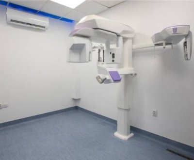
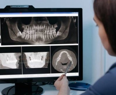
As per an article published in investigative radiology, with respect to image quality and dose, there is not much difference between cone-beam computed tomography (CBCT) and multi-slice spiral computed tomography (MSCT). But further studies showed that MSCT is a good imaging technique for soft tissue imaging while CBCT is good for hard tissue imaging. Alexander A. Schegerer, PhD, of the department of medical and occupational radiation protection, Federal Office for Radiation Protection, Neuherberg, and colleagues, brought about these results. They compared the two imaging techniques on the basis of image quality and dose.
As per Schegerer, CBCT is being used extensively for hard tissue imaging in various regions. Regarding this Schegerer said,” Cone-beam computed tomography has been established in many different clinical fields, for example, neurology, otolaryngology, cardiology, abdominal angiography, musculoskeletal imaging, and, urology. To date, there are only few reports in the literature on radiation doses and image quality for CBCT.”
The effective radiation dose for CBCT ranged from 0.35 mSv to 18.1 mSv, while local skin dose reach up to 190 mGy. “The effective dose necessary to realize the same contrast-to-noise ratio with CBCT and MSCT depended on the MSCT convolution kernel: the MSCT dose was smaller than the corresponding CBCT dose for a soft kernel but higher than that for a hard kernel”, Schegerer further added.
During the comparision of the two imaging technologies, it was found that the images spectrum taken with CBCT at tube voltages of 85/90 kV(p) were at least half of that of images measured at 103/11 kV(p) at any spiral frequency. About this Schegerer said,” As compared with MSCT using a medium-hard convolution kernel, CBCT produces images at medium noise levels and, simultaneously, medium spatial esolution at approximately the same dose. It is well suited for visualizing hard-contrast objects in the abdomen with relatively low image noise and patient dose.”
Later in the study, it was found that to optimize the patient exposure during imaging, the collimation of the radiation field must be done as per the body region to be exposed.
About us and this blog
We are a Dental Teleradiology service provider offering online interpretation and reporting of dental radiology studies like OPG, Cone Beam CT (CBCT), Dental X Ray, Sialography etc.
Request a free quote
Please let us know about your reporting requirements so that we can send you our service proposal customized to your needs.
Subscribe to our newsletter!
More from our blog
See all postsRecent Posts
- Importance of Oral and Maxillofacial Radiology in Dentistry July 1, 2021
- Dental 3D Scanning – A New Age Revolution June 30, 2021
- Looking After Your Oral Health in Festival Season June 16, 2021


