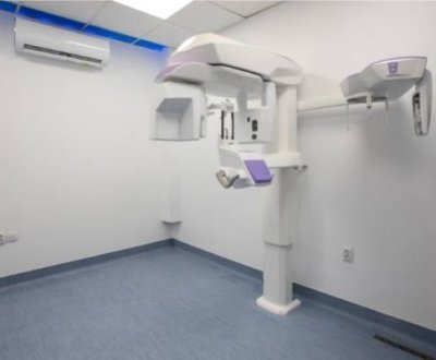
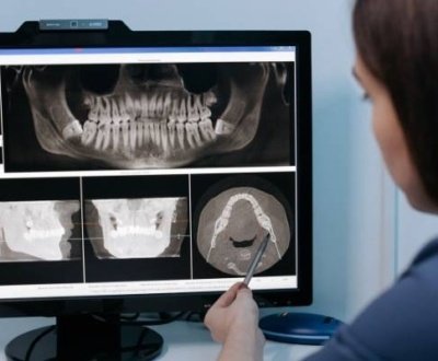
The use of CBCT for acknowledging the anatomy of the area where the implant has to be placed is widely known. In addition to that it has proved its role in detecting early implant failure. Integration of the implant with bone and postoperative complications can be diagnosed easily with the help of CBCT.
But the limitations of CBCT are similar to the other imaging modalities. This technique cannot image soft tissues such as gingiva. Also it cannot eliminate the artifacts produced by metal restorations that interfere with the correct diagnosis. In addition to this, the cost of CBCT is comparatively higher. Radiation exposure is higher than other imaging modalities. Also dentists who have little experience in interpreting the CBCT images are finding liability in reading the report.
A study conducted to judge the accuracy of CBCT in regeneration of peri-implant bone defects and determining buccal bone configuration after alveolar bone augmentation. (Clinical Oral Implants Research, Vol. 23:7, pp. 882-887) As per the study, author concluded, “ The evaluation of peri-implant bone defect regeneration by means of CBCT is not accurate for sites providing a horizontal bone width of < 0.5 mm”.
Another study conducted was regarding the use of CBCT in evaluation of changes in the buccal bone one year after implant placement. (International Journal of Oral & Maxillofacial Implants, September 2012, Vol. 27:5, pp. 1249-1257). As per this study, bony changes were qualitatively evaluated with the help of CBCT after one year of implant placement.
As per an invitro study conducted on the bovine bone, long cone technique was found to be batter when the peri-implant defect was less than 0.35mm whereas no significant difference existed between the two imaging techniques when the defect was more than 0.67mm. (Clinical Oral Implants Research, 2013, Vol. 24:6, pp. 671-678).
Another study was conducted to compare the efficacy of intraoral radiography, CBCT, panoramic radiography, and digital volume tomography (DVT) (Journal of Periodontology, July 2006, Vol. 77:7, pp. 1234-41). Out of all these techniques, DVT was found to be the best in quality, though the changes were found both in CT and DVT.
All these conducted studies pointed out to the fact that in case of post-implant cases, both the imaging modalities whether conventional radiography or CBCT are equal in the efficacy. More of such studies must be conducted to evaluate the efficacies further.
About us and this blog
We are a Dental Teleradiology service provider offering online interpretation and reporting of dental radiology studies like OPG, Cone Beam CT (CBCT), Dental X Ray, Sialography etc.
Request a free quote
Please let us know about your reporting requirements so that we can send you our service proposal customized to your needs.
Subscribe to our newsletter!
More from our blog
See all postsRecent Posts
- Importance of Oral and Maxillofacial Radiology in Dentistry July 1, 2021
- Dental 3D Scanning – A New Age Revolution June 30, 2021
- Looking After Your Oral Health in Festival Season June 16, 2021


